
Appendix B: Cattle vertebral column
Figure I
Ruminant
Click on image for larger view
Description for image - Cattle vertebral column, Ruminant
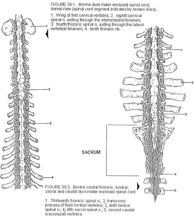
The left figure represents the dorsal view of a bovine dura mater enclosed spinal cord.
Are identified:
Number 1 is the wing of the first cervical vertebra.
Number 2 is the eighth cervical spinal nerve exiting through the intervertebral foramen.
Number 3 is the fourth thoracic spinal nerve exiting through the lateral vertebral foramen.
Number 4 is the tenth thoracic rib.
The right figure represents the bovine caudal thoracic, lumbar, sacral and caudal dura mater enclosed spinal cord. The sacrum is identified.
Are identified:
Number 1 is the thirteenth thoracic spinal nerve.
Number 2 is the transverse process of a third lumbar vertebra.
Number 3 is the sixth lumbar spinal nerve.
Number 4 is the fifth sacral spinal nerve.
Number 5 is the second caudal (coccygeal) vertebra.
Extracted from: Sisson and Grossman The Anatomy of the Domestic Animals - Volume 1
Figure II
Bovine lumbar vertebra
Click on image for larger view

Description for image - Bovine lumbar vertebra
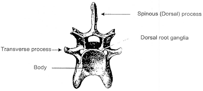
This figure represents a frontal view of a bovine lumbar vertebra. The body, transverse process, dorsal root ganglia and spinous (dorsal) process are identified.
Bovine cervical vertebra
Click on image for larger view
Description for image - Bovine cervical vertebra
This figure represents a frontal view of a bovine cervical vertebra. The body, transverse process, dorsal root ganglia and spinous (dorsal) process are identified.
Note: The Dorsal Root Ganglia may protrude from the intervertebral foramen into the space that lies between the body and transverse processes of adjoining vertebrae. The ribs are attached to shortened transverse processes in the thoracic region.
Extracted from: Sisson and Grossman The Anatomy of the Domestic Animals - Volume 1
Figure III
Bovine sacrum, ventral view
Click on image for larger view

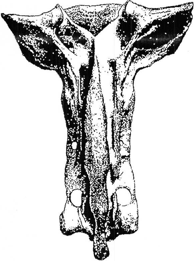
Description for image - Bovine sacrum, ventral view
This figure represents the ventral view of the sacrum of a bovine. Four arrows indicate the location of the dorsal root ganglia. The left and right wings of the bovine sacrum are identified.
Extracted from: Sisson and Grossman The Anatomy of the Domestic Animals - Volume 1
Bovine sacrum, dorsal view
Click on image for larger view
Description for image - Bovine sacrum, dorsal view
This figure represents a dorsal view of the sacrum of a bovine. No structures are identified on this figure.
Extracted from: Sisson and Grossman The Anatomy of the Domestic Animals - Volume 1
- Date modified: